Physics of Medical Scans
Positron Emission Tomography
Purpose
A PET (Positron Emission Tomography) scan is used to produce three dimensional images of the inside of the body. The scan provides information on the function of organs/tissues, along with their appearance.
PET scans are used in numerous medical investigations and to image the body. They are used to look for cancerous areas in the body or to plan operations like coronary artery bypass grafts. They are also often used for mapping brain function to identify diseases like dementia and plan brain surgery. [1]
What to expect?
In the hours leading up to the scan, you may have to alter your diet depending on what part of the body is being scanned, and why the scan is being done. Your doctor would always make you aware of this in advance.
When you arrive for the scan, a nurse will inject a radiotracer into your arm and then you will be required to quietly wait for it to be carried around the body and accumulate in certain areas, so the scanner can work effectively.
The scan itself will last up to an hour, and you will be required to lie still on a bed in the centre of a large, cylindrical scanner. After the scan, you may be asked to stay away from those who are especially vulnerable to radiation, such as babies, for a few hours. [1]
PET scans are often combined with other scans (for example CT scans) to increase the information available to doctors. In a PET-CT scan, the CT element is used to produce a high-resolution image of an area of the body, and the PET element is used to determine which cells are more active than usual. This combination is better for cancer diagnosis than a PET scan alone. [2]
For more information about PET-CT scans, click here.
Form more information about PET-MRI scans, click here.
How does it work?
A PET scanner produces images by detecting the radiation emitted by a liquid (a radiotracer) that has been injected into the body. This radiotracer collects in different organs and tissues, enabling the scan to be used to determine organ function as well as appearance.
Usually, the radiotracer used is fluorodeoxyglucose (FDG). The body accepts and transports this as it would glucose (a naturally occurring sugar), as it is structurally very similar. The radiotracer emits a small amount of radiation, which is then detected by the scanner. By analysing the areas with more and less radiation, and therefore where the radiotracer has and has not collected, it is possible to work out how well certain organs/bodily functions are working and identify any abnormalities in the body.
To find out more about how the radiotracers are made, click here.
PET scans are often used to identify cancer in the body. This is because cancerous cells metabolise (use up) glucose much faster than regular cells, so FDG concentration is higher in these cells. Therefore, PET scans are especially useful for confirmed cancer cases to monitor its spread and response to treatments.
To find out why cancerous cells use glucose faster than regular cells, click here.
Click here for a more detailed and scientifically involved explanation.
References:
[1] PET Scans. Available at: https://www.nhs.uk/conditions/pet-scan/. [Accessed 12/01/19]
[2] PET-CT Scan. Available at: https://www.cancerresearchuk.org/about-cancer/cancer-ingeneral/tests/pet-ct-scan. [Accessed 21/02/19]
[3] Huang, B. et al (2009) Whole-Body PET/CT Scanning: Estimation of Radiation Dose and Cancer Risk. Radiology. Volume 251(1), pp. 166-173.
I suffer from claustrophobia and am worried about the scan...
Unlike other scanners, PET scanners are shallow so you will never feel like you are contained in the scanner. If your scan is a combined scan (e.g. with an integrated CT scan), doctors may be able to give you a mild sedative to help you relax. [2]
I'm worried about radiation from the scan...
The level of radiation in a PET scan is low. Even a whole-body PET-CT scan leads to an exposure less than ten times the annual radiation dose from background sources [3]. For most PET scans, this is only around three times the natural annual dose. [1] This increase does marginally increase the risk of cancer in later life, so doctors will only give a scan if the clinical advantages outweigh this small risk.
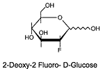
Hover your mouse here to find out why the body treats FDG like glucose.
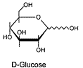
Regular glucose that cells metabolise has the structure shown left.
FDG has a very similar structure (below) but with an -OH group of atoms (a hydroxyl group) replaced by a fluorine atom.
Cells metabolise
the tracer just like glucose.
Diagram source: Seaton Hall University